Nori Hamster IFNb ELISA Kit
$461.00 – $832.00
DataSheet CoA SDS
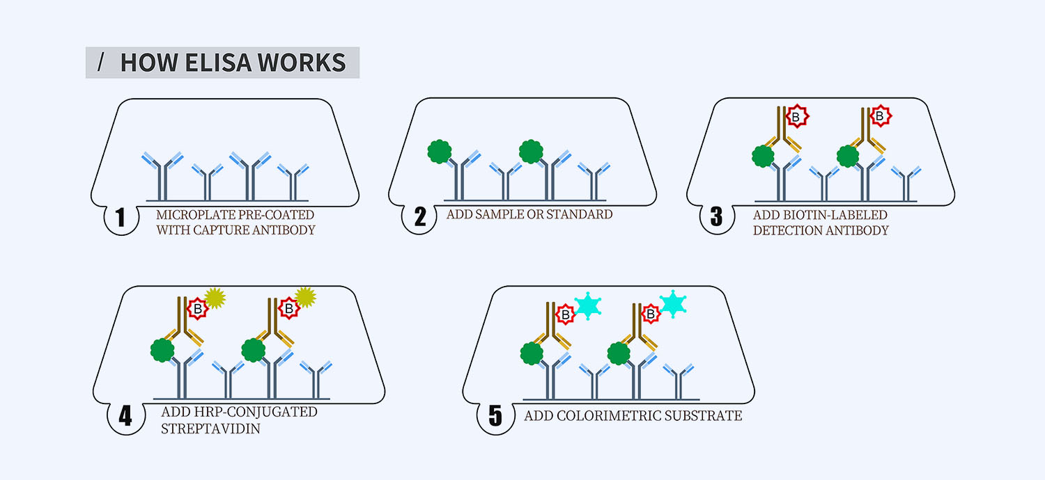
This ELISA kit is for quantification of IFNb in hamster. This is a quick ELISA assay that reduces time to 50% compared to the conventional method, and the entire assay only takes 3 hours. This assay employs the quantitative sandwich enzyme immunoassay technique and uses biotin-streptavidin chemistry to improve the performance of the assays. An antibody specific for IFNb has been pre-coated onto a microplate. Standards and samples are pipetted into the wells and any IFNb present is bound by the immobilized antibody. After washing away any unbound substances, a detection antibody specific for IFNb is added to the wells. Following wash to remove any unbound antibody reagent, a detection reagent is added. After intensive wash a substrate solution is added to the wells and color develops in proportion to the amount of IFNb bound in the initial step. The color development is stopped, and the intensity of the color is measured.
Alternative names for IFNb: Interferon beta
This product is for Laboratory Research Use Only not for diagnostic and therapeutic purposes or any other purposes.
- Description
- How Elisa Works
- Product Citation (0)
- Reviews (0)
Description
Nori Hamster IFNb ELISA Kit Summary
Alternative names for IFNb: Interferon beta,
Alternative name for hamster: Syrian hamster, golden hamster
| Assay Type | Solid Phase Sandwich ELISA |
| Format | 96-well Microplate or 96-Well Strip Microplate |
| Method of Detection | Colorimetric |
| Number of Targets Detected | 1 |
| Target Antigen Accession Number | na |
| Assay Length | 3 hours |
| Quantitative/Semiquantitative | Quantitative |
| Sample Type | Plasma, Serum, Cell Culture, Urine, Cell/Tissue Lysates, Synovial Fluid, BAL, |
| Recommended Sample Dilution (Plasma/Serum) | No dilution for sample <ULOQ; sufficient dilution for samples >ULOQ |
| Sensitivity | 5 pg/mL |
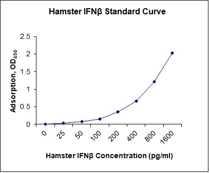
| Detection Range | 25-1600 pg/mL |
| Specificity | Natural and recombinant hamster IFNb |
| Cross-Reactivity | < 0.5% cross-reactivity observed with available related molecules, < 50% cross-species reactivity observed with species tested. |
| Interference | No significant interference observed with available related molecules |
| Storage/Stability | 4 ºC for up to 6 months |
| Usage | For Laboratory Research Use Only. Not for diagnostic or therapeutic use. |
| Additional Notes | The kit allows for use in multiple experiments. |
Standard Curve
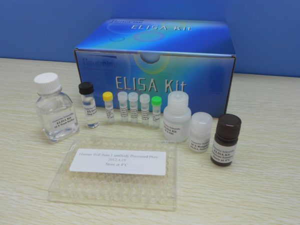
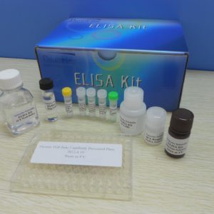
Kit Components
1. Pre-coated 96-well Microplate
2. Biotinylated Detection Antibody
3. Streptavidin-HRP Conjugate
4. Lyophilized Standards
5. TMB One-Step Substrate
6. Stop Solution
7. 20 x PBS
8. Assay Buffer
Other Materials Required but not Provided:
1. Microplate Reader capable of measuring absorption at 450 nm
2. Log-log graph paper or computer and software for ELISA data analysis
3. Precision pipettes (1-1000 µl)
4. Multi-channel pipettes (300 µl)
5. Distilled or deionized water
Protocol Outline
1. Prepare all reagents, samples and standards as instructed in the datasheet.
2. Add 100 µl of Standard or samples to each well and incubate 1 h at RT.
3. Add 100 µl of Working Detection Antibody to each well and incubate 1 h at RT.
4. Add 100 µl of Working Streptavidin-HRP to each well and incubate 20 min at RT.
5. Add 100 µl of Substrate to each well and incubate 5-30 min at RT.
6. Add 50 µl of Stop Solution to each well and read at 450 nm immediately.
Background:
Interferons (IFNs) are proteins made and released by host cells in response to the presence of pathogens such as viruses, bacteria, parasites or tumor cells, and are very important for fighting viral infections. They allow for communication between cells to trigger the protective defenses of the immune system that eradicate pathogens or tumors. IFNs belong to the large class of glycoproteins known as cytokines and are named after their ability to “interfere” with viral replication within host cells. IFNs have other functions: activation of immune cells, such as natural killer cells and macrophages; increasing recognition of infection or tumor cells by up-regulating antigen presentation to T lymphocytes; and increasing the ability of uninfected host cells to resist new infection by virus. Certain symptoms, such as aching muscles and fever, are related to the production of IFNs during infection. About ten distinct IFNs have been identified in mammals and are typically divided among three IFN classes: Type I, Type II and Type III IFN and IFN-β belongs to type I IFNs bind to a specific cell surface receptor complex known as the IFN-α receptor (IFNAR) that consists of IFNAR1 and IFNAR2 chains.[1]
In addition to the JAK-STAT pathway, IFNs can activate several other signaling cascades. Both type I and type II IFNs activate a member of the CRK family of adaptor proteins called CRKL, a nuclear adaptor for STAT5 that also regulates signaling through the C3G/Rap1 pathway.[2] Type I IFNs further activate p38 mitogen-activated protein kinase (MAP kinase) to induce gene transcription.[2] The immune effects of interferons have been exploited to treat several diseases. Interferon beta-1a and interferon beta-1b are used to treat and control multiple sclerosis, an autoimmune disorder. This treatment is effective for slowing disease progression and activity in relapsing-remitting multiple sclerosis and reducing attacks in secondary progressive multiple sclerosis.[3]
References
- de Weerd NA, et al. (2007). J Biol Chem 282 (28): 20053–20057.
- Platanias, L. C. (May 2005). Nature reviews. Immunology 5 (5): 375–386.
- Paolicelli, D. (2009). Biologics: Targets & Therapy 3: 369–376.



Reviews
There are no reviews yet.