Nori Equine TNFa ELISA Kit
$461.00 – $832.00
DataSheet CoA SDS
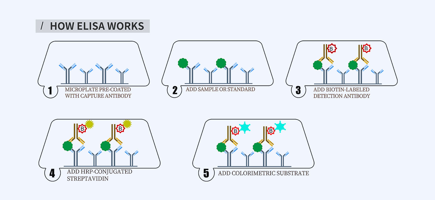
This ELISA kit is for quantification of TNFa in horse. This is a quick ELISA assay that reduces time to 50% compared to the conventional method, and the entire assay only takes 3 hours. This assay employs the quantitative sandwich enzyme immunoassay technique and uses biotin-streptavidin chemistry to improve the performance of the assays. An antibody specific for TNFa has been pre-coated onto a microplate. Standards and samples are pipetted into the wells and any TNFa present is bound by the immobilized antibody. After washing away any unbound substances, a detection antibody specific for TNFa is added to the wells. Following wash to remove any unbound antibody reagent, a detection reagent is added. After intensive wash a substrate solution is added to the wells and color develops in proportion to the amount of TNFa bound in the initial step. The color development is stopped, and the intensity of the color is measured.
Alternative names for TNFa: TNF alpha, TNF-a, tumor necrosis factor alpha
This product is for laboratory research use only not for diagnostic and therapeutic purposes or any other purposes.
- Description
- How Elisa Works
- Product Citation (25)
- Reviews (0)
Description
Nori Equine TNFa ELISA Kit Summary
Alternative names for TNFa: TNF alpha, TNF-a, tumor necrosis factor alpha
Alternative name for equine: Horse
| Assay Type | Solid Phase Sandwich ELISA |
| Format | 96-well Microplate or 96-Well Strip Microplate |
| Method of Detection | Colorimetric |
| Number of Targets Detected | 1 |
| Target Antigen Accession Number | P29553 |
| Assay Length | 3 hours |
| Quantitative/Semiquantitative | Quantitative |
| Sample Type | Plasma, Serum, Cell Culture, Urine, Cell/Tissue Lysates, Synovial Fluid, BAL, |
| Recommended Sample Dilution (Plasma/Serum) | No dilution for sample <ULOQ; sufficient dilution for samples >ULOQ |
| Sensitivity | 6 pg/mL |
| Detection Range | 31.25-2000 pg/mL |
| Specificity | Natural and recombinant equine TNFa |
| Cross-Reactivity | < 0.5% cross-reactivity observed with available related molecules, < 50% cross-species reactivity observed with species tested. |
| Interference | No significant interference observed with available related molecules |
| Storage/Stability | 4 ºC for up to 6 months |
| Usage | For Laboratory Research Use Only. Not for diagnostic or therapeutic use. |
| Additional Notes | The kit allows for use in multiple experiments. |
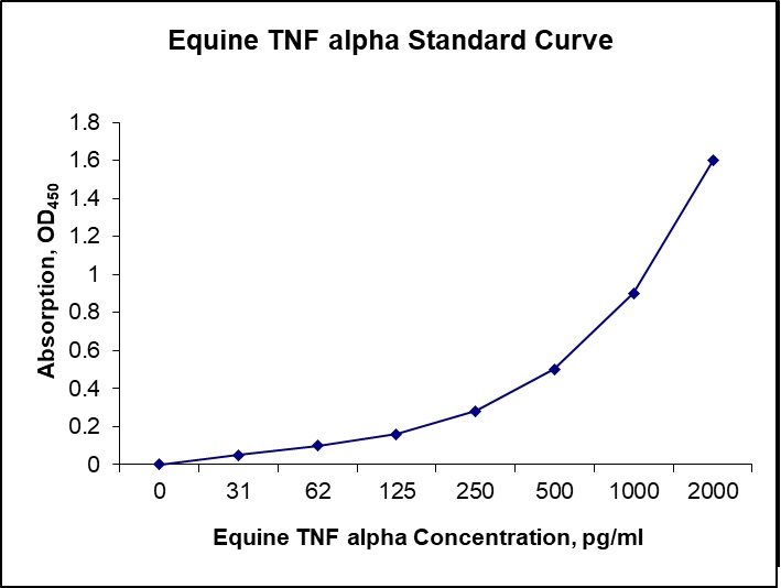
Standard Curve
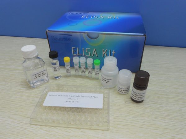
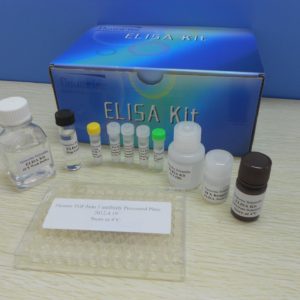
Kit Components
1. Pre-coated 96-well Microplate
2. Biotinylated Detection Antibody
3. Streptavidin-HRP Conjugate
4. Lyophilized Standards
5. TMB One-Step Substrate
6. Stop Solution
7. 20 x PBS
8. Assay Buffer
Other Materials Required but not Provided:
1. Microplate Reader capable of measuring absorption at 450 nm
2. Log-log graph paper or computer and software for ELISA data analysis
3. Precision pipettes (1-1000 µl)
4. Multi-channel pipettes (300 µl)
5. Distilled or deionized water
Protocol Outline
1. Prepare all reagents, samples and standards as instructed in the datasheet.
2. Add 100 µl of Standard or samples to each well and incubate 1 h at RT.
3. Add 100 µl of Working Detection Antibody to each well and incubate 1 h at RT.
4. Add 100 µl of Working Streptavidin-HRP to each well and incubate 20 min at RT.
5. Add 100 µl of Substrate to each well and incubate 5-30 min at RT.
6. Add 50 µl of Stop Solution to each well and read at 450 nm immediately.
Background:
TNF-α, the prototypical member of the TNF protein superfamily, is a homotrimeric type-II membrane protein (1,2). Membrane bound TNF-α is cleaved by the metalloprotease TACE/ADAM17 to generate a soluble homotrimer (2). Both membrane and soluble forms of TNF-α are biologically active. TNF-α is produced primarily by macrophages, but it is produced also by a broad variety of cell types including lymphoid cells, mast cells, endothelial cells, cardiac myocytes, adipose tissue, fibroblasts, and neuronal tissue (1). Cellular response to TNF-α is mediated through interaction with receptors TNF-R1 and TNF-R2 and results in activation of pathways that favor both cell survival and apoptosis depending on the cell type and biological context. Activation of kinase pathways (including JNK, ERK (p44/42), p38 MAPK and NF-κB) promotes the survival of cells, while TNF-α mediated activation of caspase-8 leads to programmed cell death (1,2). TNF-α plays a key regulatory role in inflammation and host defense against bacterial infection, notably Mycobacterium tuberculosis (3). TNF-α causes many of the clinical problems associated with autoimmune disorders such as rheumatoid arthritis, ankylosing spondylitis, inflammatory bowel disease, psoriasis, hidradenitis suppurativa and refractory asthma. The role of TNF-α in autoimmunity is underscored by blocking TNF-α action to treat rheumatoid arthritis and Crohn’s disease (1,2,4).
References
- Aggarwal, B.B. (2003) Nat Rev Immunol 3, 745-56.
- Hehlgans, T. and Pfeffer, K. (2005) Immunology 115, 1-20.
- Lin, P.L. et al. (2007) J Investig Dermatol Symp Proc 12, 22-5.
- Brennan, F.M. and McInnes, I.B. (2008) J Clin Invest 118, 3537-45.
Product Citation
40. Zucca E et al. (2016) Evaluation of amniotic mesenchymal cell derivatives on cytokine production in equine
alveolar macrophages: an in vitro approach to lung inflammation. Stem Cell Research and Therapy 7:137. Article
Product used and cited: Nori Equine TNFα/IL-6/TGFβ1 ELISA kits. Impact factor: 4.731.
91. Witonsky S et al. (2019) Can levamisole upregulate the equine cell-mediated macrophage (M1) dendritic
cell (DC1) T-helper 1 (CD4 Th1) T-cytotoxic (CD8) immune response in vitro? J Vet Internal Med 33(2):889-96.
Impact factor: 2.286.
Products used and cited: Nori Equine IL-6 ELISA Kit.
37. Siemieniuch MJ et al. (2016) Type of inflammation differentially affects expression of interleukin 1β and 6, tumor
necrosis factor-α and toll-like receptors in subclinical endometritis in mares. PLOS ONE 11(5):e0154934. Doi:10.137
/journal.pone.0154934. Impact factor: 2.806.
Product used and cited: Nori Equine IL-6 ELISA kit.
48. Water EVD et al. (2016) The preventive effects of two nutraceuticals on experimentally induced acute synovitis. Equine Veterinary Journal DOI:10.1111/evj.12629. Impact factor: 2.40.
Product used and cited: Nori Equine IL-6 ELISA Kit GR106001, GR106088.
81. Abass M et al. (2018) Local mepivacaine before castration of horses under medetomidine
isoflurane balanced anesthesia is effective to reduce perioperative nociception and cytokine release.
Equine Veterinary Journal, 50:733-738. Impact factor: 2.022
Produce used and cited: Nori Equine IL-6 and TNF-alpha ELISA Kit.
96. Broeckx SY et al. (2019) The use of equine chondrogenic-induced mesenchymal stem cells as a treatment for osteoarthritis:
A randomized, double-blinded, placebo-controlled proof-of-concept study. Equine Vet J 51(6): 787-94. Impact factor: 2.022.
Products used and cited: Nori Equine IL-6 and TGFβ3 ELISA Kits.
110. Hussein H et al. (2016) Cathepsin K inhibition renders equine bone marrow nucleated cells hypo-responsive
to LPS and unmethylated CpG stimulation in vitro. Comparative Immunology, Microbiology and Infectious Diseases
45: 40-7. Impact factor: 2.05.
Products used and cited: Nori Equine IL-1β, IL-6 and TNFα ELISA Kits.
126. Broeckx SY et al. (2019) Evaluation of an osteochondral fragment-groove procedure for induction of
metacarpophalangeal joint osteoarthritis in horses. Am J Vet Res 80(3):246-58. Impact factor: 1.07.
Products used and cited: Nori Equine IL-6 and TGFb3 ELISA Kits (GR106001, GR106134).
Be the first to review “Nori Equine TNFa ELISA Kit”
You must be logged in to post a review.





Reviews
There are no reviews yet.