Nori Canine CD38 ELISA Kit
$461.00 – $832.00
DataSheet CoA SDS
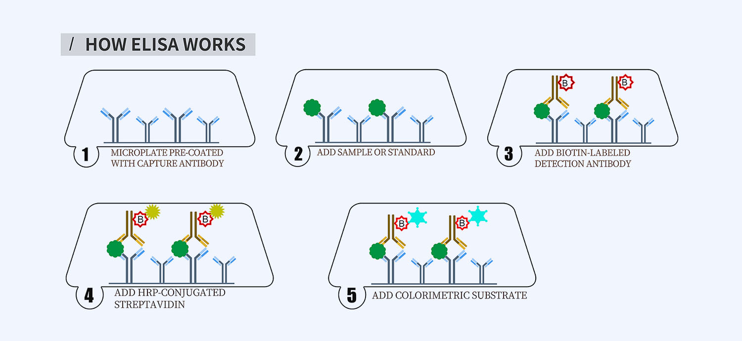
This ELISA kit is for quantification of CD38 in dog. This is a quick ELISA assay that reduces time to 50% compared to the conventional method, and the entire assay only takes 3 hours. This assay employs the quantitative sandwich enzyme immunoassay technique and uses biotin-streptavidin chemistry to improve the performance of the assays. An antibody specific for CD38 has been pre-coated onto a microplate. Standards and samples are pipetted into the wells and any CD38 present is bound by the immobilized antibody. After washing away any unbound substances, a detection antibody specific for CD38 is added to the wells. Following wash to remove any unbound antibody reagent, a detection reagent is added. After intensive wash a substrate solution is added to the wells and color develops in proportion to the amount of CD38 bound in the initial step. The color development is stopped, and the intensity of the color is measured.
Alternative names for CD38: Cluster of differentiation 38, ADPRC 1
This product is for laboratory research use only not for diagnostic and therapeutic purposes or any other purposes.
- Description
- How Elisa Works
- Product Citations
- Reviews (0)
Description
Nori Canine CD38 ELISA Kit Summary
Alternative names for CD38: Cluster of differentiation 38, ADPRC 1
Alternative names for canine: dog
| Assay Type | Solid Phase Sandwich ELISA |
| Format | 96-well Microplate or 96-Well Strip Microplate |
| Method of Detection | Colorimetric |
| Number of Targets Detected | 1 |
| Target Antigen Accession Number | NP_001003143.1 |
| Assay Length | 3 hours |
| Quantitative/Semiquantitative | Quantitative |
| Sample Type | Plasma, Serum, Cell Culture, Urine, Cell/Tissue Lysates, Synovial Fluid, BAL, |
| Recommended Sample Dilution (Plasma/Serum) | No dilution for sample <ULOQ; sufficient dilution for samples >ULOQ |
| Sensitivity | 300 pg/mL |
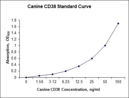
| Detection Range | 1.56-1000 ng/mL |
| Specificity | Natural and recombinant canine CD38 |
| Cross-Reactivity | < 0.5% cross-reactivity observed with available related molecules, < 50% cross-species reactivity observed with species tested. |
| Interference | No significant interference observed with available related molecules |
| Storage/Stability | 4 ºC for up to 6 months |
| Usage | For Laboratory Research Use Only. Not for diagnostic or therapeutic use. |
| Additional Notes | The kit allows for use in multiple experiments. |
Standard Curve
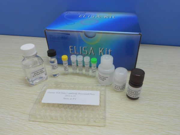
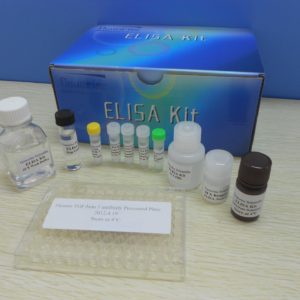
Kit Components
1. Pre-coated 96-well Microplate
2. Biotinylated Detection Antibody
3. Streptavidin-HRP Conjugate
4. Lyophilized Standards
5. TMB One-Step Substrate
6. Stop Solution
7. 20 x PBS
8. Assay Buffer
Other Materials Required but not Provided:
1. Microplate Reader capable of measuring absorption at 450 nm
2. Log-log graph paper or computer and software for ELISA data analysis
3. Precision pipettes (1-1000 µl)
4. Multi-channel pipettes (300 µl)
5. Distilled or deionized water
Protocol Outline
1. Prepare all reagents, samples and standards as instructed in the datasheet.
2. Add 100 µl of Standard or samples to each well and incubate 1 h at RT.
3. Add 100 µl of Working Detection Antibody to each well and incubate 1 h at RT.
4. Add 100 µl of Working Streptavidin-HRP to each well and incubate 20 min at RT.
5. Add 100 µl of Substrate to each well and incubate 5-30 min at RT.
6. Add 50 µl of Stop Solution to each well and read at 450 nm immediately.
Background:
CD38 (cluster of differentiation 38) encoded by CD38 gene,[1] also known as cyclic ADP ribose hydrolase is a glycoprotein found on the surface of many immune cells. CD38 functions in cell adhesion, signal transduction and calcium signaling. CD38 can function either as a receptor or as an enzyme.[2] As a receptor, CD38 can attach to CD31 on the surface of T cells, thereby activating those cells to produce a variety of cytokines.[2] CD38 is a multifunctional enzyme that catalyzes the synthesis of ADP ribose (ADPR) (97%) and cyclic ADP-ribose (cADPR) (3%) from NAD+.[3] CD38 is thought to be a major regulator of NAD+ levels,.[4][3] When nicotinic acid is present under acidic conditions, CD38 can hydrolyze nicotinamide adenine dinucleotide phosphate (NADP+) to NAADP.[3] These reaction products are essential for the regulation of intracellular Ca2+. CD38 is believed to control or influence neurotransmitter release in the brain by producing cADPR.[5] CD38 within the brain enables release of the affiliative neuropeptide oxytocin.[6] CD38 increases airway contractility hyperresponsiveness, is increased in the lungs of asthmatic patients, and amplifies the inflammatory response of airway smooth muscle of those patients. CD38 on leukocytes attaching to CD16 on endothelial cells allows for leukocyte binding to blood vessel walls, and the passage of leukocytes through blood vessel walls. The cytokine interferon gamma and lipopolysaccharide induce CD38 expression on macrophages.[7] Interferon gamma strongly induces CD38 expression on monocytes. Tumor necrosis factor strongly induces CD38 on airway smooth muscle cells inducing cADPR-mediated Ca2+, thereby increasing dysfunctional contractility resulting in asthma.[8] The loss of CD38 function is associated with impaired immune responses, metabolic disturbances, and behavioral modifications including social amnesia possibly related to autism.[9] The CD38 is a marker of cell activation and increased expression of CD38 is an unfavourable diagnostic marker in chronic lymphocytic leukemia and is associated with increased disease progression.[10]
References
- Jackson DG, Bell JI (1990). Journal of Immunology. 144 (7): 2811–5.
- Kar A, Mehrotra S, Chatterjee S (2020). Cells. 9 (7): 1716.
- Guedes A, et al. (2020). Current Opinion in Pharmacology. 51: 29–33.
- Chini EN, et al. (2002). The Biochemical Journal. 362 (Pt 1): 125–30.
- Tolomeo S, et al. (2020). Neuroscience & Biobehavioral Reviews. 115: 251–272.
- Higashida H, et al. (2019). Cells. 9 (1): 62.
- Deshpande DA, et al. (2018). Mediators of Inflammation. 2018: 8942042.
- Marlein CR, et al. (2019). Cancer Research. 79 (9): 2285–2297.
- Zambello R, et al. (2020). Cells. 9 (3): 768.
- Deshpande DA, et al. (2017). Pharmacology & Therapeutics. 172: 116–126.






Reviews
There are no reviews yet.